Home
/ Internal Thoracic Artery Relations - Internal Thoracic Mammary Artery : The crossing of the phrenic nerves with the internal thoracic arteries is seen.
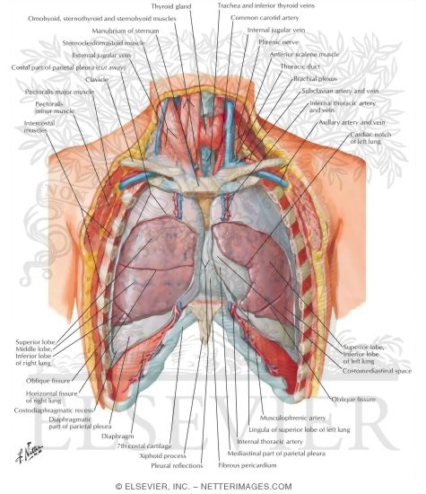
Internal Thoracic Artery Relations - Internal Thoracic Mammary Artery : The crossing of the phrenic nerves with the internal thoracic arteries is seen.
Internal Thoracic Artery Relations - Internal Thoracic Mammary Artery : The crossing of the phrenic nerves with the internal thoracic arteries is seen.. The internal thoracic artery arises from the anterior surface of the subclavian artery near its origin. First part lies medial to the anterior scalene muscle. It is accompanied by the internal thoracic vein. It also defines the range of the clearly visible, uncovered by the muscle part of the internal thoracic artery and the completeness of the muscular layer over it. Several reports have provided distinct evidence in regard to the relationship between the structural properties of the ita and cardiovascular risk factors, as previously reviewed by us 6 .
The first measurement was performed shortly after the internal thoracic artery was dissected from the chest wall and the second just prior to performing distal anastomosis to the left anterior descending coronary artery. Carotid sheath containing common carotid artery, internal jugular vein and vagus nerve infrahyoid muscles viz sternothyroid and sternohyoid ; The internal thoracic artery was studied because of its recent use in the revascularization of the myocardium in patients with coronary artery disease. Its origin, relation to the phrenic nerve and origin of the pericardiacophrenic artery were. The latter gives out the remaining anterior intercostal branches.
Https Azslide Com Download Anatomy Of The Thorax 5a4686431723ddc19fcead27 Html from Cardiac surgery october / 2017 2. Superior epigastric (rectus muscle) and musculophrenic (diaphragm) branches: It also defines the range of the clearly visible, uncovered by the muscle part of the internal thoracic artery and the completeness of the muscular layer over it. As a result it forms a unique trunk that runs up on the medial side of the artery and finishes in the corresponding brachiocephalic vein. In the event of thoracic or abdominal aortic stenosis or occlusion, the internal thoracic arteries are important potential sources of collateral blood supply via anterior anastomoses with the intercostal arteries and inferior epigastric arteries, respectively. It runs inferomedially and enters the thoracic cage deep to the clavicle and the first rib. • the first and second posterior intercostal arteries, which are given off by the superior intercostal artery (= branch of the costocervical trunk, see d a) It travels downward on the inside of the rib cage, approximately 1 cm from the sides of the sternum, and thus medial to the nipple.
The vertebral artery passes through the vertebral foramen in the cervical vertebrae and then through the foramen magnum into the cranial cavity to supply blood to the brain and spinal cord.
Internal thoracic artery is an artery which frequently used in coronary bypass operations. The measurements were made in 11 thorax dissections and morphological data of ita were obtained Superior epigastric (rectus muscle) and musculophrenic (diaphragm) branches: The crossing of the phrenic nerves with the internal thoracic arteries is seen. Carotid sheath containing common carotid artery, internal jugular vein and vagus nerve infrahyoid muscles viz sternothyroid and sternohyoid ; Enters thorax, posterior to clavicle. Internal thoracic ( mammary ) artery 1. The transversus thoracis plays an important role in heart surgery as it lies in immediate relation to the internal thoracic artery, clinically known as the internal mammary artery (ima). The internal thoracic artery, or mammary artery, supplies blood to the thymus, the pericardium of the heart, and the anterior chest wall. The internal thoracic (it) vessels (otherwise known as the thoracica interna or internal mammary vessels) are widely used as recipient vessels in autologous breast reconstruction. Value of internal thoracic artery grafting to the left anterior descending coronary artery at coronary reoperation. During the time interval between the two measurements the internal thoracic artery was immersed in a special applicator tube. Runs downwards and lateral to sternum.
Thoracic duct and phrenic nerve (on left side) posterior: It runs inferomedially and enters the thoracic cage deep to the clavicle and the first rib. The internal thoracic artery supplies the anterior body wall from clavicle to umbilicus including the sternum and the breasts. The measurements were made in 11 thorax dissections and morphological data of ita were obtained Runs downwards and lateral to sternum.
Https Azslide Com Download Anatomy Of The Thorax 5a4686431723ddc19fcead27 Html from Apex of the lung covered by the cervical pleura and supra pleural membrane. It has a close relation to the internal thoracic artery (ita), which is now accepted as a superior graft for cabg surgery 1, 2 . The vertebral artery passes through the vertebral foramen in the cervical vertebrae and then through the foramen magnum into the cranial cavity to supply blood to the brain and spinal cord. Skeletonization of bilateral internal thoracic artery is not associated with an increased risk of deep sternal wound infection. In fact, they are often preferred to the thoracodorsal (td) vessels due to their accessibility, ease of manipulation and freedom of flap placement 11, 28 . The internal thoracic artery was studied because of its recent use in the revascularization of the myocardium in patients with coronary artery disease. Internal thoracic artery is an artery which frequently used in coronary bypass operations. In most cases, the artery is left intact at its origin, with the opposite end sewn to the coronary artery below the site of the blockage.
The arterial loops are not obvious in muscles, but intercostal arteries can be seen as loops between the aorta and bilateral internal thoracic aa.
Apex of the lung covered by the cervical pleura and supra pleural membrane. The internal thoracic artery arises from the anterior surface of the subclavian artery near its origin. It travels downward on the inside of the rib cage, approximately 1 cm from the sides of the sternum, and thus medial to the nipple. First part lies medial to the anterior scalene muscle. The crossing of the phrenic nerves with the internal thoracic arteries is seen. It is interesting to note that among the first uses of the internal thoracic artery for myocardial revascularization negative clinical results were found attributed to limited blood flow (singh et al., 1973) owing to inadequate calibers (grondin et al., 1975) or other causes of reduced blood flow to the heart, like the theft of fluid from a. Several reports have provided distinct evidence in regard to the relationship between the structural properties of the ita and cardiovascular risk factors, as previously reviewed by us 6 . In fact, they are often preferred to the thoracodorsal (td) vessels due to their accessibility, ease of manipulation and freedom of flap placement 11, 28 . In the event of thoracic or abdominal aortic stenosis or occlusion, the internal thoracic arteries are important potential sources of collateral blood supply via anterior anastomoses with the intercostal arteries and inferior epigastric arteries, respectively. It has a close relation to the internal thoracic artery (ita), which is now accepted as a superior graft for cabg surgery 1,2. Superior epigastric (rectus muscle) and musculophrenic (diaphragm) branches: During the time interval between the two measurements the internal thoracic artery was immersed in a special applicator tube. It has a close relation to the internal thoracic artery (ita), which is now accepted as a superior graft for cabg surgery 1, 2 .
The latter gives out the remaining anterior intercostal branches. According to classical textbook descriptions 3,4, the fibers of transversus thoracis form At 6th intercostal space divides into: In the event of thoracic or abdominal aortic stenosis or occlusion, the internal thoracic arteries are important potential sources of collateral blood supply via anterior anastomoses with the intercostal arteries and inferior epigastric arteries, respectively. Apex of the lung covered by the cervical pleura and supra pleural membrane.
Anterior Relations Of The Heart from www.netterimages.com The internal thoracic artery, via its superior epigastric branches anastomoses with the inferior epigastric arteries, provide an important alternate blood supply if the aorta becomes occluded between the arch of the aorta and descending aorta. Cardiac surgery october / 2017 2. In addition, via the pericardiophrenic artery it supplies the mediastinum, thymus, phrenic nerves and pericardium. Thoracic duct and phrenic nerve (on left side) posterior: The internal thoracic artery arises from the anterior surface of the subclavian artery near its origin. Internal thoracic ( mammary ) artery 1. The left phrenic nerve (lpn) crosses anteriorly with the left internal thoracic artery (lita), and the right phrenic nerve (rpn) crosses posteriorly the right internal thoracic artery (rita). Internal thoracic artery is an artery which frequently used in coronary bypass operations.
It runs inferomedially and enters the thoracic cage deep to the clavicle and the first rib.
It also defines the range of the clearly visible, uncovered by the muscle part of the internal thoracic artery and the completeness of the muscular layer over it. Each of the subclavian arteries is made up of three parts, defined in relation to the anterior scalene muscle. Thoracic duct and phrenic nerve (on left side) posterior: In most cases, the artery is left intact at its origin, with the opposite end sewn to the coronary artery below the site of the blockage. The chest wall is supplied principally by the posterior intercostal arteries, which arise from the aorta, and by anterior intercostal branches arising from the internal thoracic artery: The latter gives out the remaining anterior intercostal branches. The vertebral artery passes through the vertebral foramen in the cervical vertebrae and then through the foramen magnum into the cranial cavity to supply blood to the brain and spinal cord. The internal thoracic artery, via its superior epigastric branches anastomoses with the inferior epigastric arteries, provide an important alternate blood supply if the aorta becomes occluded between the arch of the aorta and descending aorta. To see where it goes, we'll look at a dissection of the anterior chest wall by itself, seen from behind. The internal thoracic artery (previously called as internal mammary artery) then divides into the superior epigastric artery and musculophrenic artery. Its origin, relation to the phrenic nerve and origin of the pericardiacophrenic artery were. • the first and second posterior intercostal arteries, which are given off by the superior intercostal artery (= branch of the costocervical trunk, see d a) The rationale for subdividing the intercostals arteries into dorsal and ventral parts relates to the arterial bifurcation ventrally (fig.
The arterial loops are not obvious in muscles, but intercostal arteries can be seen as loops between the aorta and bilateral internal thoracic aa internal thoracic artery. At 6th intercostal space divides into: